
Rabu, 20 Maret 2013
Westgard Rule
What are the "Westgard Rules"? How do you use them?
Everything you ever wanted to know, or possibly didn't want to know, about multirule QC. Multirules are popularly known in the laboratory as "Westgard Rules." Here's the best place to find out more about them.
- What is Multirule QC
- What are the "Westgard Rules"?
- What are the "Westgard Rules"?
- What are other common multirules?
- How do you perform multirule QC?
- What is N?
- Why use a multirule QC procedure?
- Are there similar strategies for QC testing and diagnostic testing?
- Are there similar performance characteristics for QC and diagnostic tests?
- How can you use multiple tests to optimize performance?
- When should you use a multirule QC procedure?
LOOKING FOR "WESTGARD RULES" WORKSHEETS? CLICK HERE
What is a multirule QC procedure?
First, a non-technical description. When my daughter Kristin was young and living at home, she liked to party. One day when she told me she was again intending to be out late, I felt the need to exert some parental control over her hours. So I told her that if she was out once after three, twice after two, or four times after one, she was in big trouble. That's multirule control.
Kristin hates it when I tell this story, and while it isn't entirely true, it's still a good story and makes multirule QC understandable to everyone. (By the way, she turned out okay; she graduated number 1 in her class from law school and I'm very proud of her. It's also true that she has her mother's brains, which together with my persistence - or stubborness, as it's known around the house - makes a pretty good combination.) I will also have to admit that around our house it is Mrs. Westgard's rules that really count. My wife Joan hates it when I tell this part of the story, but she's put up with me for over thirty-five years and I'm now in a state of fairly stable control, so it will take a bigger deviation than this before I get into big trouble.
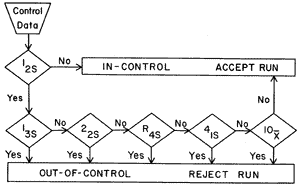
Now for a more technical description. Multirule QC uses a combination of decision criteria, or control rules, to decide whether an analyticalrun is in-control or out-of-control. The well-known Westgard multirule QC procedure uses 5 different control rules to judge the acceptability of an analytical run. By comparison, a single-rule QC procedure uses a single criterion or single set of control limits, such as a Levey-Jennings chart with control limits set as either the mean plus or minus 2 standard deviations (2s) or the mean plus or minus 3s. "Westgard rules" are generally used with 2 or 4 control measurements per run, which means they are appropriate when two different control materials are measured 1 or 2 times per material, which is the case in many chemistry applications. Some alternative control rules are more suitable when three control materials are analyzed, which is common for applications in hematology, coagulation, and immunoassays.
What are the "Westgard rules"?
For convenience, we adopt a short hand notation to abbreviate different decision criteria or control rules, e.g., 12s to indicate 1 control measurement exceeding 2s control limits. We prefer to use subscripts to indicate the control limits, but other texts and papers may use somewhat different notation (e.g. 1:2s rather than 12s) Combinations of rules are generally indicated by using a "slash" mark (/) between control rules, e.g. 13s/22s.
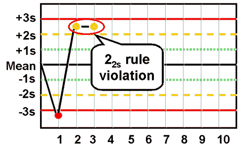
The individual rule are defined below. The "thumbnail" graphic next to a rule shows an example of control results that violate that rule. You can click on a graphic to get a larger picture that more clearly illustrates the application of each control rule.
13s refers to a control rule that is commonly used with a Levey-Jennings chart when the control limits are set as the mean plus 3s and the mean minus 3s. A run is rejected when a single control measurement exceeds the mean plus 3s or the mean minus 3s control limit.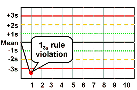 |
12srefers to the control rule that is commonly used with a Levey-Jennings chart when the control limits are set as the mean plus/minus 2s. In the original Westgard multirule QC procedure, this rule is used as a warning rule to trigger careful inspection of the control data by the following rejection rules.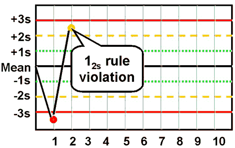 |
22s - reject when 2 consecutive control measurements exceed the same mean plus 2s or the same mean minus 2s control limit.
 |
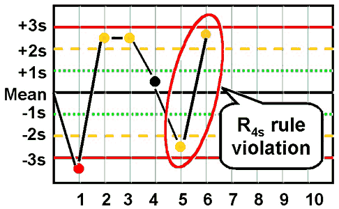
R4s - reject when 1 control measurement in a group exceeds the mean plus 2s and another exceeds the mean minus 2s.
 |
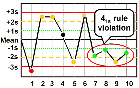
41s - reject when 4 consecutive control measurements exceed the same mean plus 1s or the same mean minus 1s control limit.
 |
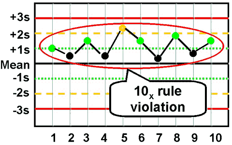
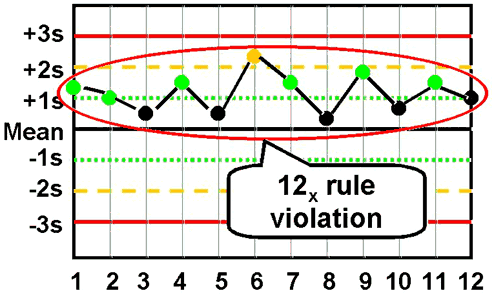
10x - reject when 10 consecutive control measurements fall on one side of the mean.
 |
| In addition, you will sometimes see some modifications of this last rule to make it fit more easily with Ns of 4: |
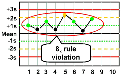
8x - reject when 8 consecutive control measurements fall on one side of the mean.
 |
12x - reject when 12 consecutive control measurements fall on one side of the mean.
 |
The preceding control rules are usually used with N's of 2or 4, which means they are appropriate when two different control materials are measured 1 or 2 times per material.
What are other common multirules?
In situations where 3 different control materials are being analyzed, some other control rules fit better and are easier to apply, such as:
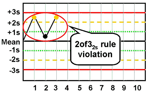
2of32s - reject when 2 out of 3 control measurements exceed the same mean plus 2s or mean minus 2s control limit;
 |
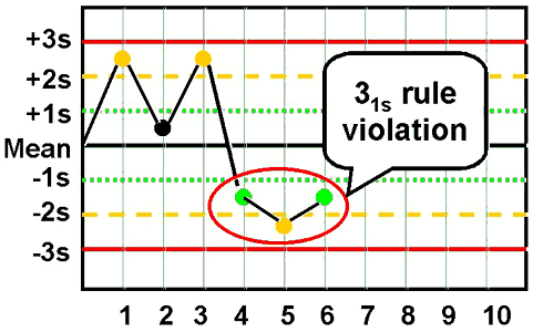
31s - reject when 3 consecutive control measurements exceed the same mean plus 1s or mean minus 1s control limit.
 |
6x - reject when 6 consecutive control measurements fall on one side of the mean.
 |
| In addition, you will sometimes see some modification of this last rule to include a larger number of control measurements that still fit with an N of 3: |
9x - reject when 9 consecutive control measurements fall on one side of the mean.
 |
| A related control rule that is sometimes used, particularly in Europe, looks for a "trend" where several control measurements in a row are increasing or decreasing [note: it is increasingly rare to see this rule in use]: |
7T - reject when seven control measurements trend in the same direction, i.e., get progressively higher or progressively lower.
 |
How do you perform multirule QC?
You collect your control measurements in the same way as you would for a regular Levey-Jennings control chart. You establish the means and standard deviations of the control materials in the same way. All that's changed are the control limits and the interpretation of the data, so multirule QC is really not that hard to do! For manual application, draw lines on the Levey-Jennings chart at the mean plus/minus 3s, plus/minus 2s, and plus/minus 1s. See QC - The Levey Jennings chart for more information about preparing control charts.
In manual applications, a 12s rule should be used as a warning to trigger application of the other rules, thus anytime a single measurement exceeds a 2s control limit, you respond by inspecting the control data using the other rules. It's like a yield or warning sign at the intersection of two roads. It doesn't mean stop, it means look carefully before proceeding.
How do you "look carefully"? Use the other control rules to inspect the control points. Stop if a single point exceeds a 3s limit. Stop if two points in a row exceed the same 2s limit. Stop if one point in the group exceeds a plus 2s limit and another exceeds a minus 2s limit. Because N must be at least 2 to satisfy US CLIA QC requirements, all these rules can be applied within a run. Often the 41s and 10x must be used across runs in order to get the number of control measurements needed to apply the rules. A 41s violation occurs whenever 4 consecutive points exceed the same 1s limit. These 4 may be from one control material or they may also be the last 2 points from a high level control material and the last 2 points from a normal level control material, thus the rule may also be applied across materials. The 10x rule usually has to be applied across runs and often across materials.
Computer applications don't need to use the 12swarning rule. You should be able to select the individual rejection rules on a test-by-test basis to optimize the performance of the QC procedure on the basis of the precision and accuracy observed for each analytical method and the quality required by the test.
What is N?
When N is 2, that can mean 2 measurements on one control material or 1 measurement on each of two different control materials. When N is 3, the application would generally involved 1 measurement on each of three different control materials. When N is 4, that could mean 2 measurements on each of two different control materials, or 4 measurements on one material, or 1 measurement on each of four materials.
In general, N represents the total number of control measurements that are available at the time a decision on control status is to be made.
Why use a multirule QC procedure?
Multirule QC procedures are obviously more complicated than single rule procedures, so that's a disadvantage. However, they often provide better performance than the commonly used 12s and 13s single-rule QC procedures. There is a false-alarm problem with a 12s rule, such as the Levey-Jennings chart with 2s control limits; when N=2, it is expected than 9% of good runs will be falsely rejected; with N=3, it is even higher,about 14%; with N=4, it's almost 18%. That means almost 10-20%of good runs will be thrown away, which wastes a lot of time and effort in the laboratory. While a Levey-Jennings chart with 3s control limits has a very low false rejection rate, only 1% or so with Ns of 2-4, the error detection (true alarms) will also be lower, thus the problem with the 13s control rule is that medically important errors may not be detected. (See QC - The Rejection Characteristics for more information about the probabilities for error detection and false rejection.)
The advantages of multirule QC procedures are that false rejections can be kept low while at the same time maintaining high error detection. This is done by selecting individual rules that have very low levels of false rejection, then building up the error detection by using these rules together. It's like running two liver function tests and diagnosing a problem if either one of them is positive. A multirule QC procedure uses two or more statistical tests (control rules) to evaluate the QC data, then rejects a run if any one of these statistical tests is positive.
Are there similiar strategies for QC testing and diagnostic testing?
Yes, a QC test is like a diagnostic test! The QC test attempts to identify problems with the normal operation of an analytical testing process, whereas the diagnostic test attempts to identify problems with the normal operation of a person. Appropriate action or treatment depends on correctly identifying the problem.
Both the QC test and the diagnostic test are affected by the normal variation that is expected when there are no problems,i.e., the QC test attempts to identify changes occurring beyond those normally expected due to the imprecision of the method, whereas the diagnostic test attempts to identify changes beyond those normally expected due to the variation of a population (the reference range or reference interval for the test) or the variation of an individual (intra-individual biological variation). The presence of this background variation or "noise" limits the performance of both the QC test and the diagnostic test.
Are there similar performance characteristics for QC and diagnostic tests?
This background variation causes false alarms that waste time and effort. These false alarms are more properly called false positives for a diagnostic test and false rejections for a QC test, but both are related to a general characteristic called "test specificity." True alarms are called true positives for a diagnostic test and are referred to as error detection for a QC test, and both are related to a general characteristic called "test sensitivity." Sensitivity and specificity, therefore, are general performance characteristics that can be applied to a test that classifies results as positive or negative (as for a diagnostic test) or accept or reject (for a QC test).
Diagnostic tests are seldom perfectly sensitive and perfectly specific! Therefore, physicians have developed approaches and strategies to improve the performance of diagnostic tests. One approach is to adjust the cutoff limit or decision level for classifying a test result as positive or negative. Both sensitivity and specificity change as this limit changes and improvements in sensitivity usually come with a loss of specificity, and vice versa.
QC procedures, likewise, seldom perform with perfect error detection and no false rejections. Laboratories can employ similar approaches for optimizing QC performance. Changing the control limit is like changing the cutoff limit, and improvements in sensitivity usually come at a cost in specificity (the 12s rule is an example). Wider control limits, such as 2.5s, 3s, and 3.5s lead to lower error detection and lower false rejections.
How do you use multiple tests to optimize performance?
Another approach for optimizing diagnostic performance is to use multiple tests. To improve sensitivity, two or more tests are used together and a problem is identified if any one of the tests is positive - this is parallel testing. To improve specificity, a positive finding from a sensitive screening test can be followed up with a second more specific test to confirm the problem - this is serial testing. Both sensitivity and specificity can be optimized by a multiple testing approach, but again these changes usually affect both characteristics.
Strategies with multiple tests can also be used to optimize the performance of a QC procedure. Multirule QC is the general approach for doing this. The objectives are to reduce the problems with the false alarms or false rejections that are caused by the use of 2s control limits, while at the same time improving error detection over that available when using 3s control limit. The multiple tests are different statistical tests or different statistical control rules, and the strategies are based on serial and parallel testing.
- False alarms are minimized by using the 12s rule as a warning rule, then confirming any problems by application of more specific rules that have a low probability of false rejection (serial testing).
- True alarms or error detection are maximized by selecting a combination of the rules most sensitive to detection of random and systematic errors, then rejecting a run if any one of these rules is violated (parallel testing).
When should you use a multirule QC procedure?
Not always! Sometimes a single rule QC procedure gives you all the error detection needed while at the same time maintaining low false rejections. This generally means eliminating the 12s rule because of its high false rejections and considering others such as 12.5s, 13s, and 13.5s which have acceptably low false rejection rates. The remaining issue is whether adequate error detection can be provided by these other single rule QC procedures. If medically important errorscan be detected 90% of the time (i.e., probability of error detection of 0.90 or greater), then a single rule QC procedure is adequate. If 90% error detection can not be provided by a single rule QC procedure, then a multirule QC procedure should be considered. In general, you will find that single rule QC procedures are adequate for your highly automated and very precise chemistry and hematology analyzers, but you should avoid using 2s control limits or the 12scontrol rule to minimize waste and reduce costs. Earlier generation automated systems and manual methods will often benefit from the improved error detection of multirule QC procedures.
To figure out exactly when to use single rule or multirule QC procedures, you will need to define the quality required for each test, look at the precision and accuracy being achieved by your method, then assess the probabilities for false rejection (Pfr) and error detection (Ped) of the different candidate QC procedures. Aim for 90% error detection (Ped of 0.90 or greater) and 5% or less false rejections (Pfr of 0.05 or less). With very stable analytical systems that seldom have problems, you may be able to settle for lower error detection,say 50%. (See QC - The Planning Process for practical approaches to select appropriate single rule and multirule QC procedures.)
For more information, see these references:
- Westgard JO, Barry PL, Hunt MR, Groth T. A multi-rule Shewhart chart for quality control in clinical chemistry. Clin Chem 1981;27:493-501.
- Westgard JO, Barry PL. Improving Quality Control by use of Multirule Control Procedures. Chapter 4 in Cost-Effective Quality Control: Managing the quality and productivity of analytical processes. AACC Press, Washington, DC, 1986, pp.92-117.
- Westgard JO, Klee GG. Quality Management. Chapter 16 in Fundamentals of Clinical Chemistry, 4th edition. Burtis C, ed., WB Saunders Company, Philadelphia, 1996, pp.211-223.
- Westgard JO, Klee GG. Quality Management. Chapter 17 in Textbook of Clinical Chemistry, 2nd edition. Burtis C, ed., WB Saunders Company, Philadelphia, 1994, pp.548-592.
- Cembrowski GS, Sullivan AM. Quality Control and Statistics, Chapter 4 in Clinical Chemistry: Principles, Procedures, Correlations, 3rd edition. Bishop ML, ed., Lippincott, Philadelphia, 1996, pp.61-96.
- Cembrowski GS, Carey RN. Quality Control Procedures. Chapter 4 in Laboratory Quality Management. ASCP Press, Chicago 1989, pp.59-79.
Jumat, 15 Maret 2013
Promosi Kesehatan
|
PROMKES
|
(47
& 54)
>WHO 1947 dan UU no. ( / 1960-PK KES
sehat yaitu kedaan sejahtera sempurna dari fiisk, mental, dan sosial yang tidak hanya terbatas pada bebas dari pnyakit / klemahan saja
>WHO 1957
sehat yaitu suatu keadaan dan kualitas dari organ tubuh yang berfungsi secara wajar dengan segala faktor keturunan dan lingkungan yang di punyainya.
>WHO 1986
the proces of enabling people to increase
control over and to improve theirnhealth
Macam-macam pendekatan yg ada dlm bina suasana
-pendekatan individu : individu2 tkoh masy. mnyebarluaskn opini positif thadap perilaku yg d perkenalkan,
cth, pemuka agama rajin 3M
-pdktn kelompok : spt pengurus RT, RW, majelis pengajian, organisasi profesi
-Pdktn Masy. umum : masy umum mbina dan mmanfaatkn radio, tv, kooran agar mjd peduli thdp apa yg d kenalkn
hal yg harus d perhatikan dl strategi promkes,
a. integrasi dg program keshtn
b. saling mnguntungkan
c. pddkn tatanan
a. integrasi dg program keshtn
b. saling mnguntungkan
c. pddkn tatanan
hub. hari kesehatn nasional 12 november 1955
dg promkes :
pada hari kesehatn nasional 12 november 1955 erat kaitannya dg merubah pola tradisional dlm promkes u/ mciptakan kmitraan sbg upaya promkes o/ pmerinthh n swasta
dg tujuan :
- mningkatkn tg.jwb sosial thdp kshtn
- mningkatkn investasi u/ pmbangunann kshtn
- mningkatkn kmitraan kshtn
- mningkatkn kmampuan perorangann pmberdayaan mash d bidang kshtn
- mningkatkan infrastruktur promkes
tujuan dari kemitraan.
1) Tujuan umum:
a. Meningkatkan percepatan
b. Efektifitas,
kuantitas dilakukan efektif atau tidak
c. Efisiensi
2) Tujuan khusus:
a. Menyamakan persepsi
dan meningkatkan pemahaman untuk mencapai Indonesia sehat 2010
b. Memperluas wawasan
tentang kemitraan
c. Mengembangkan gagasan bangkes
d. Menggalang sumber
daya (dana, tenaga, sarana)
e. Menjalin kemitraan dg bangkes prinsip kemitraan:
-kesetaraan
-keterbukaan
-saling menguntungkan
-kesetaraan
-keterbukaan
-saling menguntungkan
3 strategi dsr promkes:
-integrasi dg program kshtn
-landasan fakta
-pndekatan tatanan
-integrasi dg program kshtn
-landasan fakta
-pndekatan tatanan
. Strategi promkes
Strategi :
1.
Advokasi
Advokasi adalah :
Upaya atau proses untuk memperoleh komitmen, yang dilakukan secara persuasif
dengan menggunakan informasi yang akurat dan tepat.
2.
Bina suasana
Bina suasana adalah
upaya menciptakan opini atau lingkungan sosial yang mendorong individu anggota
masyarakat untuk mau melakukan perilaku yang diperkenalkan
3.
Gerakan pemberdayaan
masyarakat
Pemberdayaan
masyarakat di bidang kesehatan adalah upaya memampukan masyarakat sehingga
mereka mempunyai daya/kekuatan untuk hidup mandiri di bidang kesehatan
4.
Kemitraan
Kemitraan adalah
hubungan (kerjasama) antara dua pihak atau lebih, berdasarkan kesetaraan,
keterbukaan, dan saling menguntungkan.
Usaha pemerintah dalam pelaksanaan perilaku
sehat
1. Tekanan (enforcement)
adalah upaya agar masyarakat merubah perilaku
kesehatan dengan cara2 tekanan, paksaan atau koersi. Upaya ini diwujudkan dalam
bentuk UU, instruksi, dan sangsi2.
2. Edukasi
adalah upaya agar masyarakat berperilaku sehat
dengan cara persuasi, bujukan, himbauan, memberikan informasi melalui
pendidikan kesehatan.
7. 3
pendekatan dalam bina suasana
1)
Bina suasana individu ditujukan pada
individu-individu tokoh masyarakat, menyebarluaskan opini yang positif terhadap
perilaku yang sedang diperkenalkan, misalnya seorang pemuka agama yang rajin
melaksanakan 3M, yaitu menguras, menutup dan mengubur untuk mencegah wabah
demam berdarah
2)
Bina suasana kelompok ditujukan kepada
kelompok masyarakat, seperti pengurus RT, pengurus RW, Majelis pengajian,
organisasi profesi, dll
3)
Bina suasana masyarakat umum dilakukan
terhadap masyarakat umum dengan membina dan memanfaatkan media komunikasi,
seperti radio, tv, koran, dll, sehingga dapat tercipta pendapat umum,
pendekatan ini diharapkan media massa tersebut menjadi peduli dan mendukung
perilaku yang sedang diperkenalkan.
3 Prinsip dasar kemitraan
1.
Kesetaraan
·
kepercayaan penuh
·
dihargai, dihormati
·
pengakuan, kemampuan
2.
Keterbukaan
·
jujur
·
terbuka
·
tidak merahasiakan
3.
Saling menguntungkan
·
keuntungan
·
manfaat
usaha pmerintah dlm plaksanaan prilaku sehat
- preventif
- promotif
- curatif
- mlindungi diri
- aktf dlm grakan sht
the proces of enabling people to increase control over and to improve theirnhealth
- preventif
- promotif
- curatif
- mlindungi diri
- aktf dlm grakan sht
the proces of enabling people to increase control over and to improve theirnhealth
Parasitologi 1
KISI-KISI PARASITOLOGI
A.
Helmintologi
Medik
1.
Bentuk infektif &
diagnostic
a.
Lumbricoides:
larva.stad II, telur matang (dibuahi/tdk).
b.
Cacing
Tambang: larva filariform, telur dlm tinja.
c.
Filarial:
larva stad. III, mikrofilaria.
d.
F.
hepatica: metasekaria dlm tumbhn air, telur tak berembrio
e.
H.
Nana: telur berembrio.
2.
Cacing Filaria (I)
a.
Species yg ada
di Indonesia & nama penyakitnya:
Wuchereria bancrofti (nama penyakitnya
filariasis bankrofti) dan Brugia malayi (nama penyakitnya filariasis malayi).
b.
Stadium2 cacing &terdapatnya:
L1:
lambung nyamuk, L2’L3: otot toraks& kpla nymuk, L4’L5 (c.dewsa btina, jntn):
klnjar limfe manusia.
c.
Jenis2 periodisitas
& mikrofilarianya: noktuma : muncul dlm darah tepi pada malam hari. Dan
pd siang berada dlm kapiler organ2 dlm (viseral). Mikrofilaria: eosinofilik paru2 (tak bredar dlm darah),
mikrofilaria dlm peredaran darah.
d.
Hospes
definitive & perantaranya:
Definitif: manusia, perantara: nyamuk(W.
Bancrofti : Anopheles sp, Aedes sp, Culex quinguefasciatus) , (B.malayi:
Mansonia uniformis, Anopheles barbirostris).
3.
Cacing Filaria(II)
a.
Sampel &
cara diagnostic:
Sampel:darah mlm hari. Sediaan tetes
tebal u/ melihat grakan aktif microfilaria. Pnetapan spesies dgn pwarnaan
(giemsa/wright)
b.
Aspek klinik
W.Brancofti:
infeksi disbabkan c. dws/mikrofilaria, mnimbulkan limfadenopati
&limfangitis retrogard, dlm kondisi trtentu occult filariasis.
c.
Vector penularannya:
culex
quinguefasciatus, aedes sp, anopheles sp.
d.
Epidemiologi
W.Brancofti: ditemukan
dipedesaan maupun prkotaan. Trutama pdesaan, penyebaran brsifat local.
4.
Schistosoma Japonicum
a.
Stadium2 cacing:
telur,
mirasidium, sporokista I&II, serkaria.
b.
Aspek klinik: disebabkan oleh
jml c.dws yg bnyk & ektopik. Timbul klainan hati> fibrosis hati,
hepatosplenomegali & limfadenopati pd infeksi kronik. Gejala: gatal2, demam
tinggi, eosinofilia, diare, &disentri.
c.
Cara diagnosis: menemukan telur
yg khas dalam tinja/dlm jar.hati &rectum.
d.
Epidemiologi: endemic di
Sulawesi tengah, skitar danau Lindu dan Lembah Napu.
5.
Tania Solium
a.
Morfologi cacing
dewasa: cacing
dewasa: seperti pita, berwarna putih,panjang 2-4 m. tubuh terdiri dari skoleks,
leher, dan strobila, Skoleks berbentuk seperti bola dengan 4 batil isap.
Strobila tersusun oleh proglotid.
b.
Aspek klinik: Infeksi
disebabkan oleh cacing dewasa dan larvanya. Gejala klinik yang timbul
diantaranya iritasi ringan pada usus tempat perlekatan cacing, nyeri ulu hati,
sakit kepala, anoreksi, lemah, gejala abdominal samar-samar. Menyebabkan
peritonitis, obstruksi. Prognosis oleh cacing dewasa umumnua baik, prognosis
oleh sistiserkus buruk.
c.
Cara diagnosis: menemukan
telur dan cacing dewasa. Telur yang ditemukan digunakan untuk identifikasi
tingkat genus. Proglitid T. solium mempunyai cabang lateral uterus antara7-13,
T. saginata 15-20.
d.
Epidemiologi: Frekuensi tiap daerah berbeda. Berhubungan dengan kebiasaan penduduk mengkonsumsi daging babi,
adat keagamaan dan kesadaran higienik dan santasi yang kurang.
6.
Mencari alasan
a.
Untuk diagnosis
cacing tambang tidak cukup hanya dengan menemukan telur, karena dapat juga menemuka bentuk larva infektif dari hasil
biakan tinja (metoda HARADA-MORI)
b.
Pengambilan
sampel darah penderita filariasis dilakukan malam hari, karena mikrofilarianya bersifat periodesitas noktuma :
muncul dalam darah tepi pada malam hari. Dan pada siang berada dalam kapiler
organ dalam (viseral) .
7.
Gambar
diagramatik daur hidup:
a.
Filaria
(W.Brancofti)
b.
Schistosoma
Japonicum
c.
Tania
saginata
8.
Pengertian
a.
Perioditas
nokturna:
mikrofilaria terdapat di dalam darah tepi pada malam
hari.
b.
Anemia hipokhrom
mikrisiter:
Ukuran sel-sel darah merah
kecil mengandung Hemoglobin dalam jumlah yang kurang dari normal ( MCV maupun
MCHC kurang ).
c.
Mikrofilaremia: salah
satu stadium pada patogenesis filariasis bankrofti.
d.
Serkaria: bentuk
infektif cacing Schistosoma
e.
Metaserkaria: bentuk
infektif dan bentuk kista cacing Fasciola sp.
f.
Proglotida gravid:
bentuk diagnostik dalam tinja di lingkungan dari Taenia solium
g.
Emlario
heksakant:
Larva yang memiliki 6 kait dan diselubungi oleh
lapisan dalam yg disebut embrifor, yang ada di dalam telur Taenia sp
h.
Skoleks: Skoleks merupakan bagian tubuh dari cestoda
yang dilapisi kutikula, berfungsi untuk melekat pada dinding usus dan dapat
digunakan untuk mengidentifikasi spesies dalam genus Taenia.
B.
Protozoologi
Medik
1.
Pendahuluan (I)
a.
Pengertian
protozoa yang benar dan tepat, Protozoa
adalah hewan yang tubuhnya terdiri atas satu sel (monoseluler).
b.
Stadium-stadium
protozoa, Stadium kista dan stadium vegetative ( tropozoit / poliferatif).
c.
Dasar
klasifikasi protozoa, Protozoa diklasifikasikan berdasarkan
alat geraknya. Dibagi menjadi sporozoa, rizopoda, flagelata, dan ciliate
d. Macam-macam klas protozoa dan contoh spesiesnya, Sporozoa: Plasmodium vivax, P.
falciparum, P. malariae, P. ovale.
Rhizopodea : E. histolytica,
E. coli
Zoomastigophorea:
Giardia lamblia (A), Trichomonas vaginalis (A), Balantodium coli (B)
Ciliatea : Toxoplasma gondii
2.
Pendahuluan
(II)
a.
Cara reproduksi
protozoa dan contoh spesiesnya:
Seksual
: dengan pembentukan sel gamet. Contoh :
Belah
pasang : protozoa membelah menjadi dua dengan bentuk yang sama. Contoh :
amoeba, flagelata, dan ciliata.
Skizogoni
: protozoa membelah menjadi beberapa inti yang kemudian diselubungi
sitoplasma, membentuk merozoit.
Membentuk
kista : ekskistasi inti yang membelah menjadi kista. Tiap kista dapat
mengeluraka beberapa tropozoit baru. Contoh : amoeba,
Konyugasi
: penggabungan sementara.
b.
Penularan
(transmisi) protozoa:
Penularan prozoa dapat
secara langsung atau melaui makanan dan air setelah berada di luaara tubuh
hospes. Protozoa yang tidak memiliki bentuk kista penularannya melalui bentuk
tropozoitnya, dapat pula ditularkan melalui vektornya.
3.
Pendahuluan
(III)
a.
Patologi dan
gejala klinik protozoa, Infeksi terbagi menjadi
dua stadium yaitu stadium akut yang dapat berkembang menjadi stadium laten yang
menahun dan terkadang diselingi kambuh (relaps). Infeksi semula dapat berjalan
subklinik.
b.
Cara diagnosis: Menemukan parasit dalam tubuh traktus intestinalis
(misalnya amebiasis), dari bahan pemeriksaan berupa urin atau secret vagina
(trikomoniasis), dari darah dan jaringan (malaria). Sediaan dapat berupa apus
langsung, konsentrasi, pembiakan dan inokulasi, serta tes serologic pada
toksoplasmosis.
4.
E.histolytica
a.
Perbedaan bentuk
dan sifat bentuk histolytica dan bentuk minuta
Histolitika: Ukuran
20-40 mikron, Inti entameba terdapat dalam enoplasma.
Ektoplasma tampak bening dan
homogen.
Minuta: Ukuran
10-20 mikron,Inti entameba terdapat di dalam endoplasma yang berbutir
butir,Ektoplasma tidak nyata dan hanya tampak bila terbentuk pseudopodium.
b. Amebiasis intestinal: Amebiasis
usus/amebiasis kolon. Ditandai adanya radang usus besar yang disebut colitis
ulserosa. Akut : gejala jelas, tinja berlendir, bentuk histolitika mudah
ditemukan. Kronik : gejala tidak jelas, diare diselingi obstipasi, bentuk
histolitika sulit ditemukan.
c.
Diagnosis kolon
akut: Gejala diare berlangsung tidak lebih 10 kali sehari. Menemukan
bentuk histolitika dari E. histolytica dalam tinja.
d.
Diagnosis
Amebiasis hati:
Berat badan menurun, badan
lemah dan demam, anoreksi, hepatomegali. Pemeriksaan radiologic: tampak
peninggian diafragma. Pemeriksaan darah: lekositosis. Diagnosis lab : menemukan
bentuk histolitika dalam biopsi dinding abses atau aspirasi nanah abses.
Pemeriksaan serologic: hemaglutinasi tidak langsung atau tes imunodifusi.
5.
G.Lamblia
a.
Morfologi
tropozoit dan kista
Tropozoit/vegetative/proliferative
: seperti buah jambu monyet, bilateral simetrik. Anterior membulat,
posterior meruncing, dorsal melengkung.punya dua inti dan 4 flagel.
Kista
: bentuk oval, dinding halus dan tampak jelas, sitoplasma mempunyai
butir halus, yang letaknya terpisah dengan dinding kista. Kista muda punya 2
inti, kista matang punya 4 inti yang terletak di kutub.
b.
Patologi dan
gejala klinik:
Iritasi menyebabkan sekresi mukosa usus
meningkat,dapat menimbulkan gangguan absorpsi lemak. Menyerang saluran dan
kandung empedu, menyebabkan iritasi dan penebalan mukosa, penyumbatan
bilirubin. Biasanya infeksi tanpa gejala, keluhan sakit ulu hati, sakit perut
dan kembung. Kadang gejala diare, kolesistitis dan ikterus. Pada anak ditandai
enteritis akut atau kronik disertai diare. Pada infeksi lanjut kronik, gejala
utamanya masa tinja berlemak dan diselingi obstipasi. Keadaan akut ditandai
berak encer yang kering.
c.
Diagnosis:Menemukan bentuk vegetative dalam tinja encer atau
cairan duodenum. Kista ditemukan pada masa tinja yang padat.
d.
Epidemiologi: Menyebar
secara cosmopolitan terutama dari lingkungan keluarga besar. Penularan parasit
melalui makanan dan minuman atau dengan kontak langsung.
6.
T.Vaginalis
a.
Morfologi: tidak memiliki bentuk kista. Mempunyai 4 flagel anterior
dan 1 flagel posterior yang melekat pada membrane bergelombang. Sitoplasma
bergranula, aksostil dari aran anterior ke posterior. Ditularkan dalam bentuk
tropozoit.
b.
Cara infektif: Ditularkan
dalam bentuk tropozoit, melalui hubungan seksual, atau secara tidak langsung
melalui alat mandi.
c.
Patologi dan
gejala klinik,
Terjadi deskuamasi dan degenerasi sel epitel vagina.
Terdapat banayk leukosit dalam secret vagina karena seranagn leukosit. Fluor
albus. Saat melalui stadium akut gejalanya mulai berkurang. Secara klinik,
dinding vagina tampak berwarna kemerahan karena ptechiae.
d.
Cara diagnosis: Klinik
: keluhan keputihan, rasa pana, gatal pada vagian maupun vulva, sekret encer
berbusa serta berbau tidak enak. Terdapat lesi bekas garukan da hyperemia pada
vagina.
Laboratorium : menemukan parasit
dari bahan sekret vagina, uretra, prostat, dan urin.
7.
Malaria
a.
Spesies yang ada
di Indonesia:
Plasmodium vivax, P.
falciparum, P. malariae, P. ovale
b.
Distribusi
geografik: Ditemukan pada 60o LU – 32o LS. Dari daerah
rendah 400 m dpl (laut mati) – 2 600m dpl (Londiani di Kenya). Di Indonesia
penyakit malaria ditemukan tersebar dii seluruh kepulauan
c.
Morfologi dan
daur hidup:
Daur hidup meliputi fase
seksual eksogen (sporogoni) di dalam tubuh nyamuk Anopheles sp. Dan fase
aseksual (skizogoni) dalam tubuh hospes vertebrata. Fase aseksual pada manusia
terjadi dua siklus yaitu siklus eritrosit dan siklus jaringan hati (skizogoni
eksoeritrosit).
d.
Perbedaan
morfologi P.vivax dan P.Falciparum: Pada infeksi P. valciparum
hanya mengalami satu generasi aseksual dalam hati sebelu memulai siklus
eritrosit, selanjutnya siklus jaringan hati tidak dilanjutkan lagi.
Pada
P. vivax terjadi siklus eksoeritrosit terus menerus sampai berlangsung bertahun
tahun untuk melengkapi perjalanan penyakit yang berlangsung sangat lama,
sehingga sering terjadi kekambuhan.
8.
P.falciparum
a. Morfologi
Perkembangan
aseksual dalam hati hanya mengalami fase erotrosit saja, tidak mengalami fase
eksoeritrosit yang dapat menimbulkan rekurens. Skizon dapat dilihat dalam hati
setelah hari keempat setelah infeksi. Bentuk cincin (tropozoit muda) parasit
ini sanga kecil dan halus, kadang terdapat du butir kromatin yag disebut double
chromatine. Bentuk pinggir dan bentuk accole dalam satu eritrosit.
b. Malaria
cerebral
Dimulai
secaralambat atau mendadak setelah gejala awal. Gejala berupa sakit kepala,
rasa mengantuk yang disususl keadaan koma dengan pupil mengecil dan reflex
hulang atau meninggi. Menyerupai gejala meningitis, epilepsy, delirium,
intoksikasi, sangat panas dan lain lain. Gejala ditimbulkan oleh sumbatan
kapiler pada susunan syaraf pusat oleh eritrosit yang mengandung parasit.
c.
Diagnosis
Menemukan
parasit stadium tropozoit muda (bentuk cincin) tanpa atau dengan gametosit
dalam sediaan darah tepi. Pada autopsy dapat ditemukan pigmen dan parasit dalam
kapiler otak dan alat alat dalam.
d. Criteria untuk mementukan
resistensi
9.
T.Gondii
a. Hospes
dan nama penyakit
Hospes
definitive : kucing dan hewan sejenisnya. Hospes perantara: manusia. Penyakit :
toksoplasmosis konginental dan akuisita.
b. Cara
infeksi
1. Penularan
parasi ke janin terjadi secara in utero melalui plasenta (ibu mendapat infeksi
waktu hamil)
2. Pada
toksoplasma akuisita hospes memakan daging mentak atau dimasak kurang sempurna
yang mengandung kista atau proliferative parasit atau menelan ookista yang
berasal dari tinja kucing.
3. Infeksi
dari laboratorium dari sampel dengan parasit hidup.
c.
Toksoplasmosis congenital
d. Diagnosis
Cairan
serebral : perubahan tidak khas, dipastikan bila kadar protein yang ada dalam
ventrikel tinggi
Darah
: lekositisis, lekopenia, lomfositosis, monositosis, trombositopenia, dan
eosinofilia. Tokso akut dipastikan dengan menemukan tropozoit dalam biopsy
otak, sumsum tulang belakang, cairan serebrospinal, dan ventrikel.
Kelenjar
limfe yang terinfeksi menunjukkan perubahan histologik yang khas tapi parasi
jarang ditemukan.
Tes serologic yang menunjang
: teswarna Sabin-Feldman.
10.
Pengertian
a. Kista
Bentuk
infektif parasit namun tidak aktif.
b. Endoplasma
Bagian
yang berada di dalam (diselubungi) oleh membrane plasma
c.
Amebiasis ekstra intestinal
Amebiasis
yang menyebar keluar usus.
d. Inti
kosentrik
Inti
terpusat di satu sisi
e.
Ekskistasi
Proses
keluarnya tropozoit dari kista
f.
Aksostil
alat
yang memanjang berupa batang di tenga-tengah badan yang terdapat pada beberapa
flagelata
g.
Membrane bergelombang
Selaput
yang terjadi karena flagella melingkari badan parasit. yaitu sebuah membran
yang dibentuk antara sebuah flagel dan kosta pada badan parasit.
h. Skizon
Skizon merupakan produk dari skizogoni
i.
Titik-titik Schüffner
Titik-titik halus berwarna merah muda (eritrosit pecah) yang
tampak dalam eritrosit yang terinfeksi.
1.
Gambar diagramatik daur hidup:
a.
Filaria (W.Brancofti)

b.
Schistosoma Japonicum

c.
Tania saginata

Langganan:
Postingan (Atom)